- 27/April/2019

The Best Treatment for Varicose Veins - Ananthapuri Hospitals
WHAT ARE VARICOSE VEINS?
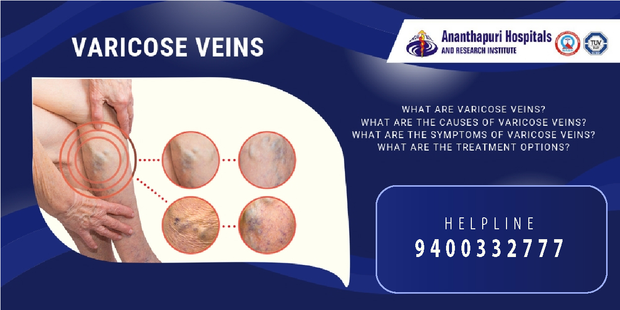
Varicose veins are visible and disfiguring venous enlargements in a part of the body, most commonly the legs. Varicose veins usually occur as a result of problems with blood flow in a large vein, causing blood to flow backwards and fluid to accumulate in the leg.
Although venous varicosities begin in childhood, they typically become visible in adulthood. Left untreated, the venous engorgement continues to expand to other branches and causes other complications, such as deep venous thrombosis (blood clot in the vein) or ulcerations in the skin overlying the venous enlargement.
WHAT ARE THE CAUSES OF VARICOSE VEINS?
Veins carry blood away from various tissues into the heart. Blood flow in the veins is determined by various forces or pressures which keep blood flow within the vessel and in one direction. These veins also have valves that keep the blood flowing in one direction.
The veins in the leg, the commonest site of varicose veins, have an extra external force – muscle activity – that helps blood flow up to the heart from the lower extremities. Any factor that causes any of the following will cause venous enlargement and varicosity:
- Impair the forward forces in the veins
- Make the vein walls more distensible or weak
- Damage the valves in the veins
- Impairs muscle activity in the legs
Some of these factors include the following:
Age
With advancing age comes weakening of the vein walls. This causes an increased distensibility of the vein, increasing the risk of varicose veins.
Genetic Disease
Some persons have a genetic failure of the venous valves. In this condition, the valves in the veins are weak and incapable of keeping blood from flowing in the opposite direction. The resultant backflow of blood causes the vein to enlarge. Genetic valvular failure is more common in women than men.
Prolonged standing or sitting
When you stand for too long, the pressure in the veins of the legs increases, since the activity of the leg muscles are reduced. This increased pressure causes the veins to distend and the valves to become weak.
Women are more susceptible to varicose veins from prolonged standing because the valves become progressively weak from the recurrent exposure to progesterone, which occurs during their menstrual period.
Pregnancy
Three factors are responsible for the high risk of varicose veins in pregnancy- 1; the hormonal changes during pregnancy causes the vein walls and valves to become more distensible and weak. 2; pregnancy is associated with an increase in maternal blood volume. This, in turn, means the veins accommodate a larger volume of blood during pregnancy. 3; as the womb enlarges, it compresses a large vein behind it, the inferior vena cava, which drains blood from all the veins in the leg and abdomen to the heart. Compressing this vein causes a backflow in the leg and abdominal veins, causing varicose veins.
Varicose veins occur more commonly among people living in industrialized nations as a result of their sedentary lifestyle and dietary choices. It is also more common among women than men at any age.
Symptoms of Varicose Veins
People with varicose veins typically present with the following symptoms:
- Leg heaviness
- Burning sensation along the length of the vein, which is aggravated by standing for a long time and relieved by walking.
- Night cramps
- Swelling of the leg
- Skin changes, with ulceration of the skin overlying the vein
- Numbness in leg
- Itching of the affected part
- Inability to walk or exercise for too long.
These symptoms are usually gradual and many people with varicose veins may not be aware of their symptoms when it begins.
Diagnosis of Varicose Veins
Varicose veins can be diagnosed by just looking at the distension of the veins. However, doctors use imaging techniques and other specialized procedures to identify the areas where the incompetence of the veins occur and where the blood flow is obstructed.
Some of these investigations include:
Duplex Ultrasound Scan
Duplex ultrasonography is the standard radiological technique for diagnosing and assessing varicose veins. This technique uses high frequency sound waves to locate the areas in the affected veins where blood flow is obstructed. Color-flow ultrasonography remains the gold standard for evaluating and diagnosing varicose veins.
Direct Contrast Venography
This radiological technique is an invasive method of diagnosis that is difficult to carry out. It involves doctors passing a catheter through a vein at the front of the foot and infusing a dye into it. The dye moves along the course of blood flow in the vein. This makes the whole length of the vein more visible on an X-ray film, and your doctor can view the areas of obstruction in the vein more clearly.
Magnetic Resonance Venography (MRV)
MRV is used to examine varicose veins in the legs and lower abdomen when other diagnostic tests are inconclusive. In addition, magnetic resonance venography may also help to exclude other causes of leg swelling.
Venous Refilling Time
Venous refilling time is a test that detects how long it takes for the blood vessels in the leg veins to fill with blood, after any muscular activity that has emptied its veins completely.
In healthy persons, the venous refilling time is about 2 minutes. In people with defects in the veins or valves, this is usually much faster.
Maximum Venous Outflow (MVO)
Doctors measure the maximum venous outflow to determine how fast blood can flow out of a congested leg vein when an external compression, such as a tourniquet placed in the thigh, is applied and removed.
The MVO is usually lower in healthy patients because the valves and vein walls are competent enough to keep blood flowing in the forward direction.
Treatment of Varicose Veins
Varicose veins are potentially dangerous because of the reflux of venous blood, which carries deoxygenated blood. If this deoxygenated blood that is rich in wastes of energy production is refluxed back to the leg tissues, it could cause the death of the leg tissue.
The main goal of treatment of varicose veins is to remove these reflux veins altogether - either by medical treatment or through surgery. Once the veins are removed, it improves blood flow.
These are the various treatment options for varicose veins:
Conservative Approach
Conservative approaches, which your doctor may recommend for treating varicose veins include:
- Regular exercises
- Weight loss
- Avoiding tight clothing
- Elevating the legs
- Avoiding long periods of standing or sitting
Another conservative method of treating varicose veins is the use of compression stockings. These stockings, as their names imply, compress the legs, adding an extra force to keep blood flowing more efficiently.
These compression stockings are available over-the-counter at pharmacy stores. There are other compression stockings available only with a doctor’s prescription.
If conservative treatment fails, your doctor may consider other therapy modalities, which may include minimally-invasive treatment or surgical treatment.
Sclerotherapy
Sclerotherapy, also called chemical sclerosis or endovenous chemoablation is the most common medical technique for treating varicose veins. Sclerotherapy is offered if ablative techniques are not suitable for you.
In this procedure, a thickening substance called sclerosant is injected into the defective vein to produce a thickened wall that eventually seals the vessel. The sealed vessel is absorbed by the body after some time and blood flow is redirected to the healthy veins. You may need more than one session of sclerotherapy to successfully treat varicose veins.
The most commonly used sclerosants for this procedure today include chemicals such as polidocanol and sodium tetradecyl sulfate. These substances have a low risk of causing allergic reactions and are less likely to cause severe adverse effects to the skin once injected.
Highly concentrated or hypertonic sodium chloride is also used as a sclerosing substance. The advantage is that sodium and chloride are natural blood contents and it has minimal risk of causing toxic effects.
In ultrasound-guided sclerotherapy, injecting the sclerosant is aided by ultrasound guidance.
Laser Therapy
This technique is also called Transcutaneous Pulsed Dye Laser and intense-pulsed light (IPL) therapy. It is an effective therapy for treating varicose veins. However, it is only used when other simpler forms of treatment, such as sclerotherapy, have failed.
This technique delivers an ablative dose of light energy to the defective vessel to damage the inner lining of the vein and cause it to seal off. The sealed off vessel gets absorbed into the body with time, and blood is redirected to the healthy veins.
The problem with laser therapy is that patients could sustain skin burns that could leave an ugly scar.
Endovenous laser therapy
Endovenous laser therapy is a minimally-invasive ablation technique, in which a laser fiber is inserted through a catheter inside the affected vein. Before starting the procedure, doctors usually inject an anaesthetic drug into the vessel to reduce the resultant pain.
The laser is fired continuously from the fiber into the vein, to damage the inner lining of the vein. This causes damage through the length of the vein, causing it to close up and be absorbed by the body. The laser is then slowly withdrawn from the vein.
Common complications of endovenous laser therapy include transient abnormal sensation (pins and needles), which resolve spontaneously.
Radiofrequency Ablation
Radiofrequency ablation is also a minimally-invasive ablative method of treating varicose veins. It is done using a specially developed radiofrequency (RF) catheter.
In this procedure, the special RF catheter is passed along the affected vein through an introducer sheath placed at the upper end of the varicose vein. The catheter is advanced until the tip reaches a certain point.
After advancing the catheter, an anaesthetic drug is injected into the vein to numb the potential pain from ablation. Thermal energy is then released through a probe guided into the vein along the whole length of the vein. The catheter is withdrawn gradually as the vein is ablated. The lining of the veins are destroyed and the vessel wall eventually seals shut. The closed vessel is absorbed by the body after some time.
Compression is important after the procedure: You will be asked to wear compression stockings for up to a week to reduce bruising and tenderness that may occur after the procedure. Also, compression stockings help reduce the risk of deep venous thrombosis after the procedure.
Radiofrequency ablation has a minimal risk of complications. Common risks include loss of sensation or abnormal sensation in the area and deep venous thrombosis. However, these risks are generally rare.
Invasive techniques can also be used to treat varicose veins. These are recommended when conservative and minimally-invasive techniques fail.
Stab Avulsion or Ambulatory Therapy
This technique involves removing short segments of the varicose veins using special phlebotomy hooks passed through tiny skin incisions made over the vein. With the patient standing, a duplex ultrasound is used to detect the locations of the defective veins before making the incisions.
The phlebotomy hooks pull out the vein until it cannot be pulled any further, detaching it from its branches. Scarring is minimal in this procedure.
Transilluminated Phlebotomy
Transilluminated phlebotomy is a relatively new procedure and its use has not been fully established. In this procedure, doctors make small incisions in the leg to remove the diseased veins with the aid of a special light called endoscopic transilluminator.
Saphenectomy
Saphenectomy is also an invasive approach to treating varicose veins and it has largely been replaced by minimally invasive modalities.
The surgeon makes a large incision in the groin to expose the junction of two great thigh veins – the saphenous and femoral veins – in the upper thigh. A stiff, flexible wire is passed into the vein to strip the defective vein and pull it out of the leg.
--------------
Ananthapuri Hospital has a team of excellent vascular surgeons who can provide expert care and a wide range of treatments for varicose veins. To book an appointment, call us at +91 9400332777 or visit our hospital at Chacka, NH Bypass, Thiruvananthapuram.
Click this link to read more about the various treatments and procedures available for varicose veins: http://www.ananthapurihospitals.com/ananthapuri_blog/image_blog/53

